Cataract
Causes, Symptoms, and Course
Cataract (Katarakt) is one of the most common causes of gradual vision deterioration in old age. Due to increasing clouding of the eye lens, vision appears as if looking through a veil – contrasts and colors decrease, and glare increases. Cataracts can already occur in children as a congenital form. Usually, however, they only develop over the course of life. Lens clouding can also result from an injury or contusion of the eye, or develop from prolonged use of cortisone (systemic and/or topical).
Affected individuals usually first notice the problem with increased light sensitivity (especially at night) and deterioration of visual performance.
In a later stage, the colors and contrasts of the surroundings are increasingly perceived as grayish and blurred. While driving, increased glare from oncoming light is noticed, or those affected are also more strongly dazzled during the day in bright light conditions.
Due to the simultaneous increasing thickening of the natural lens, its refractive power increases, so that paradoxically, with advancing cataract, a previously existing farsightedness may decrease and an existing nearsightedness may increase. It is therefore not unusual that some patients temporarily regain the ability to read without glasses and that prescription values change at relatively short intervals.
Severe visual impairments up to blindness are the consequence of an untreated cataract in advanced stages. However, it no longer needs to come to that nowadays.
At our Eye Center Prof. Koss and Colleagues in Munich, we offer you modern diagnostics, personal consultation, and a gentle cataract surgery according to current medical standards – centrally located at Nymphenburger Straße 4.
What is Cataract?
Cataract refers to the clouding and hardening of the natural eye lens. As a result, light is no longer clearly directed onto the retina, and visual quality continuously decreases.
Cataracts usually develop due to aging but can also be caused by:
- congenital forms (rare)
- eye injuries or contusions
- prolonged cortisone use
- metabolic diseases
Typical Symptoms of Cataract
Many patients notice the changes gradually at first. Common complaints include:
- Many patients notice the changes gradually at first. Common complaints include:
- blurred or “foggy” vision
- increasing glare sensitivity, especially at night
- poorer vision with backlighting
- colors appear grayer, contrasts weaker
- frequent changes in prescription strength
- uncertainty while driving
Surgical Treatment of Cataract
Fortunately, the vision loss that has already occurred can be reversed through surgery.
If you are impaired in your daily life by lens clouding, you should consider surgery. Without surgery, the visual impairment will continue to progress.
When treating cataracts, there are essentially two questions:
1. How should the lens be operated on?
Surgery is advisable as soon as the lens clouding affects daily life, for example when:
- Reading
- Driving
- Working at a screen
- Recognizing faces or details
Without surgery, the cataract will continue to progress
2. Which lens can and which lens should be implanted?
During cataract surgery, the natural lens is replaced by an individually calculated artificial lens.
Available lens types:
Monofocal Lenses (Standard)
- optimal vision at one distance
- usually distance vision without glasses possible
- reading glasses often still necessary
Toric Lenses
- ·Correction of corneal curvature (astigmatism)
EDOF Lenses (Extended Depth of Focus)
- very good vision at distance and intermediate range
- minimal glare phenomena in the evening
Multifocal Lenses
- · Goal: maximum independence from glasses
- · not suitable for every patient
We will advise you thoroughly and together select the medically best solution.
Secondary Cataract (Posterior Capsule Opacification)
Secondary cataract (posterior capsule opacification) refers to the clouding of the support apparatus (the so-called capsule) after a cataract surgery, which can occur with a time delay in up to 30% of cases after the cataract operation.
This is typically treated with the so-called Nd:YAG laser capsulotomy. However, the surgeons at our eye center are also experienced in treating more complex variants of secondary cataract, which may need to be treated with a minimally invasive procedure.
Diagnostics
What Examinations Are Performed Before Cataract Surgery?
Before cataract surgery, the eyes must be thoroughly examined. This includes determining your prescription, measuring your visual acuity with and without glasses, measuring intraocular pressure, measuring corneal curvature, measuring the axial length of the eye, calculating the artificial lens, and microscopic examination of the anterior and posterior eye segments. This is necessary to determine the best lens for you.
We always recommend that our patients have the optical measurement performed due to its greater accuracy. For patients with statutory health insurance, however, this must be billed as a private additional service.
Under certain circumstances, some of these examinations may already have been performed by another ophthalmologist.
Two measurement methods are available for measuring the eye:
- Measurement with ultrasound
and the much more accurate method, which is used by us in 99% of cases:
- Optical measurement with the OCT-based Anterion
Ultrasound measurement is part of the surgical services covered by statutory health insurance; however, the more accurate optical measurement method is not.
Must Contact Lens Wearers Take a Wearing Break?
Yes! Since contact lenses rest directly on the cornea, they can influence the shape of the cornea. Due to this change in corneal shape, the measurement accuracy for calculating the artificial lens can be compromised.
If a wearing break is taken, the cornea returns to its natural shape. Hard contact lenses, including gas-permeable ones, should not be worn for at least 3 weeks before the measurements. With hard contact lenses, your visual acuity may fluctuate during this wearing break due to the still unstable corneal shape. Measurements should only be performed once these fluctuations no longer occur – even if this takes longer than 3 weeks.
How Accurate Are the Measurement Methods for Calculating the Artificial Lens?
The calculation of the power of an artificial lens is very accurate for most people. Nevertheless, the result after surgery may not correspond to the desired value. During the healing phase after surgery, the artificial lens can slightly change its position within the eye and move minimally forward or backward on the visual axis. The extent of these shifts is not the same for everyone, which is why the actual refractive power of the eye after surgery may differ from the target.
After cataract surgery, it may therefore be possible that glasses are needed for optimal near or distance vision, or in cases of higher astigmatism.
If the deviation from the desired value is significant, however, exchange of the artificial lens can be considered. Likewise, a correction with laser or with supplementary lenses that are additionally inserted into the eye can be performed.
Particularly strongly farsighted or strongly nearsighted individuals have the greatest risk that the achieved value deviates from the calculated value.
Also for people who have already undergone LASIK or other refractive procedures, the measurement and calculation of the artificial lens is more difficult.
IOL MASTER OR ANTERION
To determine the optimal artificial lens, it is important to precisely measure the patient’s eye before cataract surgery. The goal of this preliminary examination is to achieve the best possible visual acuity after the operation.
Typically, the ultrasound method is available for the measurement. However, this conventional method only measures the eyeball length.
The internationally recognized IOL Master or the anterior segment OCT (Anterion) enables a painless and non-contact laser measurement. With this optical biometry, not only the eyeball length but simultaneously also the corneal radius and anterior chamber depth are captured. With the data obtained, the probability of achieving the best possible surgical result increases.
Biometry with the IOL Master or Anterion is not included in the statutory health insurance benefit catalog. This examination must therefore be billed privately.
Corneal Topography
The keratograph is an instrument for capturing and evaluating corneal topography. A Placido disc (ring system with concentrically alternating black and white rings) is projected onto the anterior corneal surface, the ring-shaped reflection images are captured with a video camera and analyzed by a computer system using Fourier analysis.
A significant advantage compared to ophthalmometer measurement lies in the number of measurement points. In ophthalmometer measurement, only a few measurement points are captured (two central, four peripheral). Keratographs capture 10,000 to 30,000 measurement points depending on the device, resulting in a detailed profile of the cornea. Different display modes can be selected. Corneal topography can be displayed numerically, color-coded, or as a three-dimensional surface map.
Originally, the keratograph was developed for corneal surgery. It has since also found widespread use as a preliminary examination for cataract surgery or LASIK procedures, as well as in contact lens fitting.
OPTICAL COHERENCE TOMOGRAPHY (OCT)
OCT is the worldwide standard in the diagnosis of retinal diseases, particularly of the macula, and in diseases of the optic nerve, e.g., in glaucoma.
It has become the most important instrument worldwide in the diagnosis of macular diseases and therefore represents the essential examination method for the assessment of AMD, macular edema, macular hole, or a so-called macular pucker.
- The principle is based on measuring the reflection of a light beam sent into the eye and converting it into a visible image through digital technology. The resolution is 10 micrometers.
- The procedure is painless and completely harmless.
- For several years now, we have also offered so-called OCT angiography. This allows the blood flow of multiple retinal layers in the macula to be visualized non-invasively (i.e., without dye injection via the vein).
OCT is not included in the statutory health insurance benefit catalog. This examination must therefore be billed privately.
Treatment
Cataracts cannot be treated with eye drops, medications, or visual aids. The only way to treat cataracts is surgery (cataract surgery). In this procedure, the body’s own lens is replaced by an artificial lens (intraocular lens, IOL). This lens then typically remains in the eye for a lifetime (unless it dislocates).
Cataract surgery does not, unfortunately, eliminate other visual impairments caused by glaucoma, diabetes, or macular degeneration.
Cataract surgery is the most commonly performed surgical procedure. In Germany alone, this operation is performed more than 600,000 times per year. Thanks to the many years of experience of our ophthalmologists and the modern technology available at our own surgical center and the Herzog Carl Theodor Eye Clinic, we can perform this surgery to the most modern standards. We perform this operation several thousand times per year.
Cataract Surgery in Munich with Prof. Koss and Colleagues
The only effective treatment for cataracts is cataract surgery, in which the clouded lens is replaced by an artificial lens (intraocular lens, IOL).
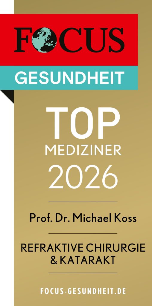
Our eye center has been continuously ranked among the practices awarded in multiple categories on the FOCUS Top Physicians List (Gold) since 2018, including in the areas of cataract, retina, and glaucoma.
The surgery is performed in our own state-of-the-art surgical center, directly connected to the eye center at Nymphenburger Straße 4.
Parking facilities are available in the immediate vicinity for family members and accompanying persons – for a relaxed organization on the day of surgery.
Course of Cataract Surgery
The surgery is almost always performed under local anesthesia, either with topical (drop) anesthesia or through an injection placed next to the eye. You will not feel the injection because a light sedation ensures that you do not notice the anesthetic injection.
Cataract surgery is typically:
- outpatient
- painless under local anesthesia
- completed within approximately 10–20 minutes
Just a few days after the procedure, vision improves significantly. The first follow-up examination usually takes place the following day.
Modern Surgical Methods
Depending on the findings, different methods may be considered:
- Ultrasound phacoemulsification (proven standard)
- laser-assisted cataract surgery as a gentle option for selected patients
Which method is medically appropriate will be discussed individually with you.
The Procedure in Three Steps:
Once you have rested briefly, you can leave our outpatient surgery center or the eye clinic and head home with your accompanying person. After the surgery, there may occasionally be a slight scratching or stinging sensation in the eye. However, most patients find this only mildly bothersome, and it disappears after a short time. Just a few days later, visual acuity has improved significantly and you can resume your normal daily activities. Maximum visual acuity is usually achieved within the next few days or weeks.
YAG Capsulotomy
YAG capsulotomy, also known as “secondary cataract opening,” is performed using the so-called YAG laser (Yttrium-Aluminum-Garnet laser). Newly proliferating cells migrate from the equatorial area into the central visual axis within the capsular bag, causing foggy, milky vision again. This opacity can be permanently opened by the YAG laser in a single session. After local anesthesia, a contact lens is placed on the cornea for better focusing of the laser beams. This procedure is low-risk and can be performed on an outpatient basis.
Lens Models
Through a thorough preliminary examination, we aim to select an artificial lens with you that can deliver the best optical result.
For most people, the best possible optical correction can already be achieved with a so-called monofocal lens.
For special visual defects, e.g., higher astigmatism, or individual wishes, e.g., the desire for freedom from glasses, only so-called lenses with additional functions can lead to the desired goal.
Lenses with additional functions are not always covered by statutory health insurance. Should a lens with additional function be the appropriate therapy for you, we will of course inform you about the expected costs including the costs for the laser-assisted surgical method. We also recommend that privately insured patients coordinate with their health insurance in advance.
HOYA Vivnex

- Function: clear vision at one set distance (usually distance).
- Benefits: very high image quality and contrast sensitivity; a good option for patients who frequently drive at night.
- Limitation: reading glasses are usually still necessary.
- Suitable for: patients focused on achieving the best possible distance vision.
- Examples / available models: HOYA Vivinex (yellow, aspherical), Alcon Clareon® Monofocal, Rayner RayOne Monofocal.
Monofocal lenses are covered by statutory health insurance. With single-focus lenses, your vision can be set to a specific distance.
- If the lens is set so that you need no or only a weak prescription for good distance vision, you will very likely need reading or progressive glasses for near vision. Conversely, patients who prefer to read without glasses and are set for this distance can then only achieve good distance vision with distance glasses. In the pre-operative consultation, we will work with you to find the best solution for your specific needs.
- With spherical monofocal lenses, occasional blurriness may occur in dim light conditions.
- Corneal curvatures (astigmatism) cannot be compensated with this lens.
Toric Lenses

- Function: additional correction of regular corneal curvature (astigmatism).
- Benefits: better distance vision without cylinder glasses and fewer residual errors.
- Examples / models: HOYA Vivinex Toric, Alcon Clareon® Toric, RayOne Toric, 1stQ AddOn® Toric.
EDOF Lenses (Extended Depth of Focus)

- 1stQ Liberty²
- Function: extended depth of focus from distance to intermediate range (e.g. screen, dashboard).
- Benefits: highly practical for everyday life, with a low tendency toward halos compared with classic multifocal lenses.
- Suitable for: patients who spend a lot of time at screens, active patients, and those wanting a high degree of independence from glasses.
- Examples / models: Alcon Vivity®, TECNIS PureSee™, RayOne EMV, Hoya Impress.
Multifocal / Trifocal Lenses

- Function: multiple focal points enable vision at distance, on the computer, and at near range (distance, intermediate, and near).
- Benefits: the greatest possible reduction in dependence on glasses after surgery; often very good independence from reading glasses.
- Limitations: possible glare or scatter effects at night; a longer adaptation period after surgery.
- Examples / models: Rayner Galaxy, 1stQ Liberty.
Toric Multifocal Lenses

- These lenses additionally combine the multifocal optical function with correction of existing regular astigmatism. They are advantageous for significant astigmatism (> −1.25 cyl) and for patients seeking independence from glasses at distance, intermediate, and near range.
Add‑On / Piggyback Lenses

- Thin supplementary lenses placed in front of an already implanted artificial lens (when the initial operation result is unsatisfactory). They can be removed again and are a good option for fine-tuning.
These lens types are currently available to us:
II. Lenses with Additional Functions
1. Toric Lenses for Correcting Corneal Astigmatism (Spherical and Aspherical)
When there is irregularity in the corneal structure, this is called corneal curvature resulting in astigmatism. This causes tilted images and blurred vision.
With the help of toric lenses, regular astigmatism can be corrected during cataract surgery. The other option to address corneal astigmatism is to make relaxing incisions at specific locations in the cornea outside the central visual zone, thereby changing the architecture of the cornea. However, the optical result is not as good as correction with toric lenses.
With toric lenses as well, the desired distance setting is determined before surgery.
Through precise calculation of the lens and corresponding positioning of the lens within the eye, this target value should be achieved as accurately as possible.
- Only with this lens can regular corneal curvature be corrected.
- With larger corneal astigmatisms, good vision without glasses at the desired distance can only be achieved with this lens.
Toric lenses are not reimbursed by statutory health insurance. The higher price of this lens and increased medical effort must therefore be billed to the patient as a private additional service.
2. Blue Filter Lenses
This yellow-tinted intraocular lens protects the retina from the short-wave rays of the blue light spectrum. Short-wave blue light is more energetic than longer-wavelength light and is considered a damaging factor in the development of macular degeneration.
- Blue filter lenses offer increased protection for your retina, as short-wave components of light are filtered out.
- Vision appears more natural.
- Color perception becomes more harmonious. Colors are perceived as “warmer” since the “cold” blue component is filtered out.
3. Bifocal Lenses with EDOF Principle
An EDOF lens refers to bifocal intraocular lenses that have an enhanced depth of focus (Extended Depth Of Focus). This achieves very good vision at distance and intermediate distance, such as kitchen counter, PC, or car dashboard.
Reading vision is usually not possible, unless you are suitable for a so-called blended vision concept. The interesting aspect of this type of lens with additional function is that it has almost no side effects (glare, etc.) in the evening.
These most modern artificial lenses have been used by us in Munich since their introduction in 2019 and are very popular among our patients, also due to our specific experience and expertise.
4. Multifocal Lenses (Trifocal Lenses)
A multifocal lens allows distance vision and can partially or completely replace the eye’s ability to focus at near distance, so that after successful surgery, glasses can largely be dispensed with.
- Multifocal lenses compensate for nearsightedness and farsightedness as well as presbyopia.
- The lens works with three focal points, projecting two focus points onto the retina. The brain can adapt within a certain time, sometimes only after several months, and can then distinguish between the two focus points.
- Depending on the interaction between your eye and the multifocal lens, blurriness may occur between the two focus points (at 60-80 cm distance, the so-called PC range).
- After surgery, you will no longer need glasses in many everyday situations.
- Due to the necessary adjustment period, multifocal lenses are not suitable for everyone. Avid readers often still need additional reading glasses.
- At night, disturbing scatter and glare effects may occur while driving. Anyone who drives frequently should be aware of this.
5. Toric Multifocal Lenses
With astigmatism, the advantages of a multifocal lens can be significantly reduced. To counteract this, toric multifocal lenses that also correct astigmatism are used for corneal curvatures from -1.25 cyl diopters.
90-95% of patients can do without glasses thanks to multifocal or toric-multifocal lenses. 5-10% of operated patients will continue to need visual aids for driving at night and for reading long or very small texts.
6. Aspherical Lenses
Aspherical lenses have optical advantages when the pupil becomes larger under certain light conditions, e.g., when driving at night.
- The special composition of their surface with a flatter curvature at the edge gives the aspherical lens advantages over the spherical lens.
- Only aspherical lenses can compensate for the so-called spherical aberration that becomes relevant with a wide pupil.
- However, they should not be used after previous hyperopic LASIK.
- For our IOL models, there is no additional charge for you here.
7. Piggyback Lens (Add-On)
If an artificial lens has already been implanted and your glasses bother you, a spherical, toric, or multifocal piggyback lens can provide relief. An additional, very thin lens is placed in front of the existing artificial lens to compensate for any remaining blurriness in near or distance vision.
- Blurriness in near and distance vision that persists even after a previous procedure can be corrected.
- If needed, the lens can be removed minimally invasively.
- more information
8. Fine-Tuning with Laser
If the desired result has not yet been achieved through lens implantation, we can fine-tune with a special laser procedure (excimer laser). Through the so-called bioptics procedure, an optical correction can be made, and our excimer laser (the most modern and newest in Munich) allows a diopter correction in one second!
Interesting fact: Did you know…?
After a thorough preliminary examination and discussion of all options, over 80% choose an individually adapted additional service, whether for the lens or the surgical method.
Health is priceless and you only have 2 eyes!
The most important thing! We are happy to advise you on the large and promising range of artificial lenses – this is our mission! You as the patient decide together with us, not we for you! Through our philosophy and especially our experience with EDOF lenses, you can rely on our recommendation prior to surgery.
We also examine and treat so-called second opinions for externally operated patients with artificial lenses. However, this is usually a private medical service.
Ultrasound / Nanopulse Method
Lens Removal by Ultrasound or Nanopulse Laser Method with Cetus
During cataract surgery, the body’s own clouded lens is first fragmented/liquefied and then removed. Until now, this was achieved using ultrasound treatment, which has been established worldwide for over 40 years.
During the treatment, energy is released that helps liquefy the clouded lens. However, this energy also affects other structures within the eye. During lens removal, this can lead to damage to cell layers.
The NanoLaser method differs fundamentally in that there is no or hardly any heat development and the energy is targeted specifically at the lens body. As a result, the endothelial cell layer is significantly less stressed.
More information here
Additional advantages of the method include:
- short surgical duration
- surgery with single-use instruments
- atraumatic surgery
- no electrical connection
The method is particularly recommended for eyes that already suffer from a pre-existing condition (such as endothelial dystrophies, glaucoma, age-related macular degeneration, etc.).
Have we sparked your interest in the gentler surgical option? Feel free to inquire at our practice at any time.
Our Financing Options for Cataract Surgery
How Financing Works
Who Can Use the Financing?
Financing is available to everyone who:
is of legal age
has their primary residence in Germany
has no negative SCHUFA entry
has a regular income or receives a pension
If needed, partners or parents can be registered as second loan recipients.
FAQ
Cataract Surgery
Frequently asked questions about cataract
What is Cataract?
Cataract refers to the clouding and hardening of the natural eye lens. As a result, light is no longer clearly directed onto the retina, and visual quality continuously decreases.
Which artificial lenses are available?
Depending on your visual goals and eye findings, we offer various lens options, including:
· monofocal lenses (standard)
· toric lenses for corneal curvature
· EDOF lenses with extended depth of focus
· multifocal premium lenses for independence from glasses
We will advise you thoroughly on which lens is medically appropriate.
How do I know if I have cataracts?
Typical signs of cataract include blurred vision, increasing glare sensitivity (especially at night), declining contrasts, and frequent changes in prescription strength. A reliable diagnosis is made through an ophthalmological examination.
When is cataract surgery necessary?
Surgery is recommended as soon as the deterioration in vision affects daily life – for example when driving, reading, or working at a screen. The timing is determined individually together with you.
How does cataract surgery work at Prof. Koss and Colleagues?
The surgery is performed on an outpatient basis and is usually painless under local anesthesia. The procedure typically takes only 10–20 minutes. Visual acuity improves significantly within just a few days.
Do you operate in your own surgical center in Munich?
Yes. We perform cataract surgery in our own state-of-the-art surgical center directly at our location at Nymphenburger Straße 4. Parking is available in the immediate vicinity for accompanying persons.
What is special about EDOF lenses?
EDOF lenses enable very good vision at distance and in the intermediate range (e.g., screen, dashboard) with simultaneously minimal glare phenomena in the evening.
Prof. Koss and Colleagues were among the first centers in Munich to adopt EDOF lenses early and build extensive experience with them.
Will I still need to wear glasses after surgery?
That depends on the chosen lens. With standard lenses, reading glasses are usually necessary. Premium lenses (e.g., EDOF or multifocal) can significantly reduce dependence on glasses.
What is secondary cataract?
Secondary cataract is a later clouding of the lens capsule that can occur months or years after cataract surgery. Treatment is performed simply on an outpatient basis with an Nd:YAG laser.
Does health insurance cover the costs?
Cataract surgery is generally covered by both statutory and private health insurance. Additional costs may arise for premium lenses or extended measurement procedures. We clarify all costs transparently in advance.
Why Prof. Koss and Colleagues in Munich?
Our eye center has been continuously listed on the FOCUS Top Physicians List (Gold) since 2018 – including in the areas of cataract, retina, and glaucoma. We combine modern technology with high surgical expertise and personal care in the center of Munich.
How can I schedule a cataract consultation appointment?
You can reach us directly at our eye center:
Prof. Koss and Colleagues Nymphenburger Straße 4, Munich Phone: 089 18 35 11